 |
| Angiosperm Life Cycle |
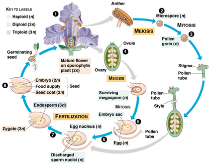
The word "angiosperm" comes from the Greek words for "vessel" and "seed" and translates roughly as "enclosed seed". In part, angiosperms (the flowering plants, phylumAnthophyta) are defined by the fact that their seeds are enclosed by an ovule. The life cycle of an angiospermis defined by the formation of the seed and its development to a full-grown plant which, in turn, produces seeds.
Angiosperms are vascular plants with flowers that produce seeds enclosed in an ovule—a fact that is recognized as the angiospermy condition.
Reproductive Flower Parts
In general, angiosperms have a floral axis with four floral parts, two of which are fertile. At the receptacle, or tip, of the axis there is an ovule-bearing leaf structure known as the carpel. The ovule or ovules can be found inside the pistil. Three portions compose the pistil: the ovary, the style, and the stigma, where the pollen usually germinates.
  |
The mature ovule consists of the placenta, the integuments that are modified leaves that cover the entrance to the embryo sac, the micropyle, and the chalaza. These latter two parts of the ovule complement each other in their positions and functions.
While the micropyle receives and guides the pollen tube, the chalaza relates to the vascular supply of the ovule, nutrition, and support. The stamens, which are often composed of the filament and sporangia sacs that make up the anther, surround the pistil. Stamens carry the male gametes, and the pistil carries the female gamete needed for sexual reproduction.
It is believed that the great diversity and adaptability of the angiosperms is related to the presence of a unique reproductive cycle. This cycle consists of an alternation of generations and the production of a pair of spores on two types of sporophylls: microspores (which become male gametophytes) and megaspores (which become female gametophytes).
Male Gamete Development
The angiosperm reproductive cycle begins with the process of microsporogenesis, or microspore formation. The stamen consists of a filament and the anther, also known as the microsporangium. Inmost of the cases, the anther consists of four pollen sacs, or locules.
Within each locule, the archesporial cell develops through mitosis and extends as a row of cells throughout the entire length of the young anther. These mitotic cell divisions generate the anther wall, which is made up of several cell layers, the outermost of which transforms itself into the epidermis. The layer of cells belowthe epidermis is known as the endothecium.
During anther development, the endothecial cells acquire thickenings whose function is related to anther opening and pollen release. The innermost layer of the anther wall is the tapetum,whose primary function correlates with the nourishment of the young pollen and the deposition of the exine, a coating of the pollen grain.
As development proceeds, the sporogenous cells located below the tapetum transform into microsporocytes. These microsporocytes will undergo meiosis, and tetrads (units of four) of microspores will form.
Shortly after their formation, the tetrads separate into individual microspores. Each microspore is haploid, and often it will enlarge and separate from the tetrad, becoming sculptured by the deposition of sporopollenin and other substances that will turn into the ornamented surface of the pollen grain.
The second phase of pollen development is known asmicrogametogenesis. Themicrospore is the first cell of the gametophytic generation, the cell that generates themature pollen. The single-nucleus microspore develops into the male gametophyte before the pollen is released.
This developmental process occurs through two or three unequalmitotic divisions of the nucleus and subsequent cytokinesis (cell separation). The two daughter nuclei and cells differ in size and in form.
The larger cell represents the tube cell and nucleus,while the smaller cell represents the generative cell and nucleus. At maturity, the grain can be shred in two or three nucleate conditions. When the anther opens, the mature male gametophytes or pollen grains will be disseminated and ready for germination.
Female Gamete Development
The ovule (female sex organ) consists of two opposite ends: the micropyle, where the integuments come together, and a more distant end, where the ovular tissue is more massive. This part is also known as the chalaza, and it lies directly opposed to the micropyle.
The mature ovule is composed of three layers: the outer integument; the inner integument; and, underneath the integuments, the nucellus. During ovular development, one cell lying below the nucellar epidermis changes into a primary archesporial; this will divide to form the primary parietal cell and primary sporogenous cell.
The primary sporogenous cell functions as the megaspore mother cell, which divides meiotically, originating four haploid megaspores. In the majority of angiosperms, three of the megaspores will degenerate, and only the chalazal one will develop into the megagametophyte (embryo sac).
After the completion of the embryo sac stage, a series of cellular events occurs, ending with the formation of the mature embryo sac, ready for fertilization by the male gametes. The chalazal megaspore enlarges and undergoes threemitoses, giving rise to eight haploid cells. The mature megagametophyte consists of two groups of four cells located at both ends of the embryo sac.
The result is three antipodals at the chalazal end: the egg apparatus (consisting of the egg and two synergids at the micropylar end) and the polar nuclei. These two cells, present at both ends, usually fuse before pollination, and during fertilization they form the primary endosperm nucleus.
Pollination
The plant reproductive structures are now ready for the union of male and female gametes or fertilization, which eventually will produce a seed with a viable embryo and cotyledons. Before that step takes place, however, the pollen must be transferred from the anther to the stigma. Biotic agents (such as birds, insects, or mammals) or abiotic agents (such as wind or water) can accomplish this transfer process, known as pollination.
After landing on the stigma, pollen tubes will emerge through the grain apertures if the environ- ment is high in humidity. Successful germination of the pollen in the stigma requires nutrients. In most plants, growth of the pollen tube lasts between twelve and forty-eight hours, frompollen germination to fertilization.
Pollen germination starts with pollen-tube initiation, elongation, and penetration of the stigmatic tissue. During this period intense metabolic activity takes place, for the tube must synthesize membrane material and cell wall for growth and expansion. Simultaneously, at its tip the tube carries the vegetative cell nucleus, fol- lowed by the germinative cell.
Angiosperms have evolved complex breeding systems that ensure they will be pollinated by their own species. Today it is recognized that two pollination syndromes exist: self-pollination and cross-pollination. In self-breeding species, the pollen comes from the anther of the same flower.
In cross-pollination (or outcrossing) species, the pollen comes from the anthers of a different flower or even a different plant of the same species. In these plants, incompatibility in the stigma guarantees that only pollen from other flowers will germinate.
Fertilization
The union of one sperm with the egg is known as fertilization. However, several developmental processes in the vegetative and germinative cells prepare the two sperms for a process known as double fertilization. A mitotic division of the germinative cell generates the spermcells. This process that can take place on the growing pollen tube or inside the pollen grain.
In a growing pollen tube, the vegetative nucleus disintegrates and the sperm cells will take the lead and enter the embryo sac for successful fertilization. Usually, the interactions between the pollen grain and the pistil ensure that the sperm cells will often reach the micropyle of the ovule.
Once the spermreach themicropyle, the growth of other tubes stops. In the embryo sac (female gametophyte), four cells are located at themicropylar side.Of those four, the first pair that the spermcells will encounter are the synergids.
One of these is always bigger than the other and carries the filiform apparatus, a structure resembling hairs that degenerates after pollination and before fertilization. The synergids act as chemical attractants to the pollen tube, which penetrates the synergids via the filiform apparatus and then releases the two sperm cells.
One of the sperm cells will fuse with the egg, producing the zygote; the other sperm cell will fuse with the primary endosperm nucleus, generating the endosperm. The remaining cells of the female gametophyte are the antipodals; they usually degenerate after fertilization has taken place.
Seed and Fruit Formation
Once fertilization has occurred, the ovule will go through a series of metabolic steps ending with the formation of the seed and the fruit. The recently created zygote transforms into amulticellular and complex embryo that has two well-defined polar ends: the radicle, or primary root, and the embryonic apical meristem with the first leaves.
After successive mitosis, the mature endosperm usually grows close to the embryo and may provide nutrients needed for germination. The integuments will undergo further transformation, replication, and elongation and will become the seed coat—of variable texture, consistency, and colors, depending on the type of plant.
In general, after pollination or during fertilization, the ovary undergoes a series of physiological changes regulated by synchronized hormonal and genetic alterations that will modify the size of the parenchyma cells and its sugar and organic acids contents.
This process turns the ovary into fruit—in many cases familiar as the edible fruits familiar in human diets. The fruit provides nourishment for the seed until it ripens and drops to the ground, where the next stage in the life cycle begins.
Germination, Seedling Development, and Maturation
Seeds are released from the fruit in a large variety of ways that have evolved to ensure the survival of species. Whether ingested by mammals and passed through their feces to the ground, borne by wind on feathery "wings", or simply falling from rotting fruit that has abscissed and dropped from the plant, the seed must next undergo a process called germination, in which the embryo enclosed in the seed begins its growth. The embryo develops a hypocotyl (root axis) and a fleshy part known as the cotyledon; inmonocots there is one cotyledon, in dicots, two.
Germination requires certain conditions, such as the softening of the seed coat, moisture, and adequate warmth, to occur. During germination, the hypocotyl begins growing downward to become the root; the cotyledon(s) will develop into the shoot, stems, and leaves.
The process of germination results in the sprouting through the ground’s surface of the seedling, which will develop into the mature plant with flowers. The cycle then begins again.